The last two decades have seen a major revolution in the treatment of varicose veins and venous reflux disease—and we may be on the brink of the next big shift. This is what Mark S Whiteley writes for Venous News, in an overview of the developments that have taken place in this field since the emergence of endovenous interventions introduced in the last 20 years, and the preceding techniques that made these possible.
In the 1890s, Friedrich Trendelenburg introduced the idea that visible varicose veins were caused by underlying truncal valve dysfunction—and to cure that, he introduced the Trendelenburg ligation. Previous to this, treatments had been aimed at the visible varices alone. The following century showed little advance in varicose veins treatment, apart from studies showing that stripping the great saphenous vein was superior to ligation alone and that the treatment of incompetent perforators helped to heal venous leg ulcers. In contrast, the last 20 years or so have seen a massive increase in the advancement of treatments for varicose veins and venous reflux disease.
Although most people point to the introduction of endovenous surgery as being the major turning point, all of the new advances actually stem from the development of venous duplex ultrasonography in the mid-1980s and early 1990s. It was only because this noninvasive imaging modality that allowed us to see the venous function in real-time became widely available that our understanding of venous disease leapt forward.
The ability to identify venous reflux, different reflux patterns, the differences between passive (diastolic) reflux that everyone understands and active (systolic) reflux that many doctors struggle with, and the size of target veins, has shaped many of the new approaches to varicose veins treatment. Moreover, the identification of venous reflux in leg varicose veins arising from pelvic veins has revolutionised the concept that varicose veins can be thought of as a problem isolated to the lower limb.
However, to think that everyone has reached the same conclusions from the advent of venous duplex ultrasound would be wrong. Whereas some have used the technique to improve understanding and hence results, most doctors just use it to identify which truncal vein they are going to treat, ignoring complex patterns, perforators or pelvic venous reflux. It is not surprising that randomised trials of different treatment modalities are not conclusive, if they only concentrate on treating incompetent truncal veins and ignore the other causes of varicose veins.
The introduction of venous duplex ultrasonography split the venous world into two main factions. Unfortunately, a great many doctors who “do” varicose veins as a job and do not attend conferences or read around the subject are unaware of this huge divide. In the English-speaking world, most doctors practice ablative surgery. Some still ligate and strip, although most now have moved on to some form of thermal ablation. Other nonthermal techniques ablative techniques are becoming more widely used.
Nevertheless, a large number of doctors remain unaware of the haemodynamic approach to varicose veins and venous reflux disease, championed by the conservative haemodynamic correction of venous insufficiency method (CHIVA). Often called “saphenous sparing surgery”, advocates of this approach present series where the results have been demonstrated to be comparable with stripping. Whereas those of us who treat all of the reflux pathways would regard such results as suboptimal, the randomised studies that have been performed where surgeons ignore perforator vein reflux and pelvic vein reflux, appear to show stripping as equivalent to the thermal ablation techniques. As such, the haemodynamic approach may begin to gain some traction.
The endovenous revolution
Following on from venous duplex ultrasonography, the biggest revolution in the treatment of varicose veins was the invention of successful endovenous thermal ablation. At the end of the 1990s, catheter-bansed radiofrequency ablation and endovenous laser ablation prove to be successful, causing endovenous surgery to take off. Not only did these endovenous thermal techniques destroy the vein, but the catheters were introduced under ultrasound control into the distal vein and passed proximally, without the need for open surgery in the groin.
This minimally invasive approach allowed the development of tumescent anaesthesia. With truncal ablation and phlebectomies being possible under tumescent anaesthesia, true “walk-in, walk-out” ambulatory surgery became possible for the treatment of varicose veins. More than just a new technique of treating veins, this allowed vein centres to be set up outside of hospitals, that could concentrate on ambulatory venous surgery.
In fast succession, treatment of incompetent perforators was developed in 2001 using the transluminal occlusion of perforator (TRLOP) technique (“reinvented” in America in 2007 as percutaneous ablation of perforator surgery or PAPS), steam vein sclerosis and several different radiofrequency and laser devices became available for leg veins.
All of these thermal ablation devices require tumescence because of the heat generated during treatment. This has led to the investigation and development of nonthermal, and therefore non-tumescence placed ablation techniques. In 1985, a patent was granted to allow detergent sclerotherapy fluids to be mixed with gas to make foam. This was then taken on by a British company hoping to replace surgery with a chemical ablation technique.
Although it has become clear in the last decade that foam sclerotherapy works well in small veins within walls, medium and long-term results have been poor in truncal veins, which are larger and have thicker walls. As such, although foam sclerotherapy is an essential technique to be used by any doctor providing venous treatments, it has been shown to have comparatively poor results when used as a sole treatment modality.
To improve sclerotherapy results, the endovenous mechanochemical ablation catheter (MOCA, Clarivein) was developed to traumatise the venous wall mechanically and to allow sclerosant to penetrate deeper. Research has shown that this increases cell death within the vein wall, improving the long term ablation over foam sclerotherapy alone. Finally, in the non-thermal nontumescent area, cyanoacrylate glue is being injected intravenously with good results in medium-term studies. This appears to use a different mechanism, as the vein wall is not ablated in the same way as it is with the previously described techniques. However, patient satisfaction is high and clinical results are very good.
Pelvic venous reflux
Over the last decade, pelvic venous reflux and pelvic congestion syndrome have become increasingly recognised as part of the varicose vein disease profile. Although we started investigating and treating this in 2000, it has largely been ignored by the venous community until more recently.
Amazingly, many of the lessons learned in the 1990s for leg varicose veins are having to be re-learned in pelvic veins. In the 1990s, doctors realised that examining varicose veins with venography, particularly supine, is suboptimal compared to venous duplex ultrasonography, where the patient was semi-erect or erect. However, many now use CT or MRI to examine pelvic veins in a supine patient! With approximately 20% of female patients with leg varicose veins having a major contribution from pelvic vein reflux, and 3% of males, it is now impossible to offer a full varicose vein service unless pelvic veins are assessed and provision for treatment is made as part of the service.
So what does the future hold for varicose vein treatment?
Undoubtedly, new endovenous devices will appear. Indeed, I have just performed the first endovenous microwave treatment in Europe. This technique has all the advantages of endovenous laser and radiofrequency ablation, but without some of the drawbacks of both. However, it is still an endovenous thermal technique requiring tumescence.
One of the most exciting new treatments for varicose veins and venous reflux disease is HIFU: high intensity focused ultrasound. This new technique has only recently been presented at meetings, so only the principles and very earliest results are known. However, HIFU has been used in other clinical scenarios for non-invasive tissue ablation and so the probability that it will be successful in veins is high.
By externally focusing ultrasound to cause ablation at one specific point targeted internally, HIFU is a truly non-invasive technique, a quantum leap forward from minimally invasive techniques. By being able to externally target specific venous areas, those of us interested in vein research will be able to explore whether ablation of all venous reflux is required, or whether a haemodynamic “CHIVA” approach can be used successfully to target specific areas of venous reflux. By being able to use the same equipment to compare strategies, we should be able to identify the optimal way of treating veins: whether it be ablation, haemodynamic or a combination of both.
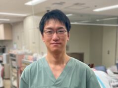
Mark S Whiteley is a consultant venous surgeon, a visiting professor at the University of Surrey in Guildford, UK, and founder of The Whiteley Clinic, with three centres in the UK.


What would the treatment differences be for these two camps of varicose vein surgeries?