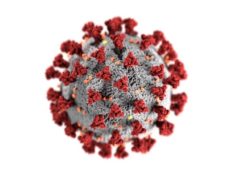
Joseph A Caprini considers venous thromboemolism (VTE), patient risk assessment, and therapeutic challenges in the wake of the COVID-19 pandemic, concluding that “accurate thrombosis risk assessment for all coronavirus patients is advised”.
Emergence of the coronavirus worldwide represents a pandemic of enormous proportion. The problem was first reported in December 2019 in Wuhan, China.1 The number of infected patients increased rapidly to more than 60,000 within the next two months. Human airway epithelial cells were used to identify a novel coronavirus called Covid-19. It was linked to pneumonia which was associated with mortality of 11.5%.2 Coronavirus pneumonia patients who died were found to have elevated D-dimer and fibrin degradation product levels (FDP) on admission and in late stages of the disease prolonged prothrombin (PT) and partial thromboplastin times (PTT). Criteria for disseminated intravascular coagulation (DIC) were found in (15) 71% of non-survivors and only 0.6% of survivors.1 Chronic diseases including cardiovascular disease, malignancy, respiratory diseases and kidney and liver abnormalities were found in 41% of the patients. During the late stages of the disease, fibrinogen and antithrombin levels were decreased along with markedly increased D dimer and FDP levels. Sepsis and consumptive coagulopathy were frequently seen.
It was postulated that these coagulation changes were based on a cascade of events beginning with inflammation and sepsis releasing inflammatory cytokines which caused increased circulating thrombin reflected in the D-dimer levels. The response to these changes was activation of the fibrinolytic system resulting in an elevation of FDP. This process involving inflammation activates coagulation resulting in increased circulating thrombin generation. Fibrinogen is converted into fibrin that coats bacteria and viruses to physically entrap them and prevent their dissemination.
The increased thrombotic state can manifest as a variety of clinical presentations. The patient may exhibit evidence of a hypercoagulable state with development of clinical VTE. DIC may evolve into consumptive coagulopathy, and as the clotting factors are used up, the PT and PTT will become prolonged. This chain of events may become impossible to manage due to bleeding, and all resources must be brought to bear, including stopping or lowering the dose of anticoagulant drugs. Transfusion of blood, platelets, or plasma products may be necessary, as well as a trial of anti-fibrinolytic drugs. Extracorporeal membrane oxygenation (ECMO) as a last resort has been used selectively.3
In a retrospective case series from Italy, involving 1591 coronavirus patients they found that 99% of 1300 patients with available respiratory support data required respiratory support, 88% required intubation and noninvasive ventilation was necessary in 11%. The ICU mortality was 26%.4
Coagulation changes representing various stages of hypercoagulability due to the presence of inflammation, sepsis and/or pneumonia are not the only pathophysiologic issues in these patients. Most Covid-19 hospitalised patients are at high risk of VTE as a result of principles proposed in 1856 by Virchow, a brilliant German pathologist. He introduced the concept known as Virchow’s triad describing three factors associated with development of venous thrombosis.5 These include venous stasis, endothelial injury, and hypercoagulability. The risk is highest when all three factors are present.
Patients who are at bed rest, particularly due to hypotension, shock, coma, sedation or when mechanical ventilation is necessary, are at risk. Marked reduction of blood flow from the legs due to inactivity occurs. This phenomenon, known as venous stasis, results in pooling of venous blood while arterial inflow is not altered. Eventually veins become engorged with blood and this results in cracks in the venous lining (endothelial injury). This process exposes the blood to sub-endothelial collagen, triggering clotting as the blood is exposed to this foreign surface. Finally, hypercoagulability exists in the calf leg veins compounded by waste products from normal muscle metabolism that become trapped and further stimulate clotting. Compounding this increased clotting activity is the individual patient’s degree of infection, sepsis, or pneumonia.
A thorough thrombosis risk assessment is particularly important in the comprehensive treatment of these coronavirus patients. The Caprini score is a comprehensive history and physical examination involving 40 elements. The weight of each element present (risk factor) (e.g., 1,2,3,5) contributing to a thrombotic event is totalled to produce a final score. This score is plotted against the clinical incidence of VTE and a nonlinear increase in clinical events with increasing score has been observed. More than 5 million patients in more than 150 clinical trials have documented this association.6
Coronavirus patients may be assessed with less complex scores but the Padua and Improve scores do not record family history of thrombosis or past obstetrical complications that may indicate the patient is a carrier of anticardiolipin antibodies or a beta2 glycoprotein abnormality.
The Caprini score can classify patients into low-risk, high-risk, or very-high-risk. Low-risk patients (scores 0–4) should not receive anticoagulant prophylaxis because it does not further lower the incidence of thrombosis, only increases bleeding complications.7 It should be noted that this principle does not apply to coronavirus patients due to their high baseline thrombotic risk. In surgical patients without the virus, high-risk patients (scores 5–8) benefit from a course of anticoagulation of at least 7 to 10 days, and very-high-risk patients (scores 9+) benefit from anticoagulant prophylaxis for up four weeks.8
Patients admitted with coronavirus are all at increased risk of thrombosis and benefit from anticoagulant prophylaxis. These patients should receive anticoagulant prophylaxis with low molecular weight heparin (LMWH) or unfractionated heparin (UFH) unless there is increased risk of bleeding. Mechanical methods are relied upon in patients where anticoagulation is not suitable. The presence of respiratory involvement automatically dictates LMWH prophylaxis because it has previously been shown to result in a 75% reduction of VTE events.9
The purpose of scoring is to identify very high-risk individuals with multiple risk factors present before the onset of the viral infection. It has been reported by Tang that 41% of patients have existing chronic disease.(1) Common examples include patients with a family history or past history of VTE, morbid obesity with a BMI over 35, or existing thrombophilia. Obese individuals frequently suffer from sleep apnea, hypertension, diabetes requiring insulin, and cardiovascular disease. It is recommended that these individuals along with those with a score of 8+ be given twice the normal dose of anticoagulation. There are no randomised trials to substantiate this practice; however, the tendency of these high-risk patients to develop fatal and nonfatal thrombotic events plus the additional thrombotic risks associated with the virus appear to justify this approach.
The Caprini score is a dynamic instrument and needs to be updated as complications during hospitalisation occur. Rescoring of the patient during hospitalisation as complications develop may push patients with lower scores into the very high-risk group. At this point then, doubling the anticoagulant prophylaxis should be considered. Increasing the score by three points occurs when patients develop a positive d-dimer. Some investigators use therapeutic anticoagulation for those with 3x or more elevation of the d-dimer levels. There is a one-point increase when inadequate ambulation occurs. The definition of ambulation has been suggested by the data to mean that walking 30 feet at one time decreases the incidence of VTE by 50%.10 Unfortunately going to the bathroom does not qualify as ambulation using these criteria.
A final Caprini score should be completed before discharge and those with a score of 8+ including those with a positive d-dimer may benefit from 6 weeks of anticoagulation facilitated by use of one of the new oral anticoagulants. Coumadin should be avoided due to the personal contact required for monitoring.
Haematologic changes signalling severe disease and a poor prognosis include one or more of the following: elevated D dimer, elevated fibrinogen or FDP, prolonged PT or PTT, and thrombocytopenia.
Some investigators have suggested screening patients who develop these changes with ultrasonography to detect subclinical thrombosis. Point-of-care ultrasound screening (POTUS) using a hand-held instrument at the bedside is advised. Limited examination including two-point ultrasound involving the femoral and popliteal veins is a practical approach. Others feel that scanning should be reserved for those with clinical indications including leg pain, swelling, etc.
In conclusion, accurate thrombosis risk assessment for all coronavirus patients is advised. Thrombosis prophylaxis is indicated for most patients and traditional anticoagulants help prevent most fatal thrombotic events. Appropriate VTE screening for select very-high-risk patients based on haematologic or clinical criteria is also advised. An aggressive approach for patients exhibiting clinical or laboratory features associated with DIC may be lifesaving.
References
- Tang N, Li D, Wang X, Sun Z. Abnormal coagulation parameters are associated with poor prognosis in patients with novel coronavirus pneumonia. J Thromb Haemost. 2020;18(4):844-7.
- Zhu N, Zhang D, Wang W, Li X, Yang B, Song J, et al. A Novel Coronavirus from Patients with Pneumonia in China, 2019. N Engl J Med. 2020;382(8):727-33.
- Rice TW, Wheeler AP. Coagulopathy in critically ill patients: part 1: platelet disorders. Chest. 2009;136(6):1622-30.
- Grasselli G, Zangrillo A, Zanella A, Antonelli M, Cabrini L, Castelli A, et al. Baseline Characteristics and Outcomes of 1591 Patients Infected With SARS-CoV-2 Admitted to ICUs of the Lombardy Region, Italy. Jama. 2020.
- Virchow RKL. Gessamelte Abhandlungen zur wissenschaftlichen Medizin. Frankfurt am Main Meidinger 1856:514-5.
- Golemi I, Adum JPS, Tafur A, Caprini J. VTE prophylaxis using the Caprini score. Disease-a-Month. 2019;65:249-98.
- Pannucci CJ, Swistun L, MacDonald JK, Henke PK, Brooke BS. Individualized VTE Risk Stratification Using the 2005 Caprini Score to Identify the Benefits and Harms of Chemoprophylaxis in Surgical Patients: A Meta-analysis. Ann Surg. 2017;265(6):1094-103.
- Cassidy MR, Rosenkranz P, McAneny D. Reducing postoperative VTE complications with a standardized risk-stratified prophylaxis protocol and mobilization program. J Am Coll Surg. 2014;218(6):1095-104.
- Turpie A. Thrombosis Prophylaxis in the Acutely Ill Medical Patient- Insights from the Prophylaxis in MEDical patients with ENOXaparin (MEDENOX) Trial.pdf. Am J Cardiol. 2000;86(suppl):48-52.
- Amin AN, Girard F, Samama MM. Does ambulation modify VTE risk in acutely ill medical patients? Thromb Haemost. 2010;104(5):955-61.
Joseph A Caprini is a senior clinician educator at the Pritzker School of Medicine at the University of Chicago in Chicago, USA. He is also an Emeritus physician at NorthShore University HealthSystem in Evanston, USA.


